


Advanced Technology
We utilize the latest available technology to better our diagnostic and treatment capabilities. Whether it is digital imaging or advanced screening technology, our practice is on the cutting edge of eye care. Putting such commitment to technology raises the level of service provided at our Idaho Falls eye care center to what you deserve.
Schedule Your Exam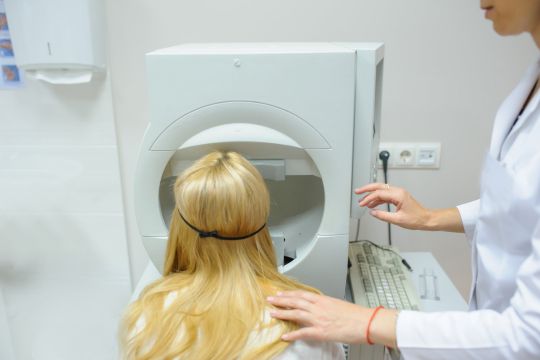
Visual Fields
A visual field test measures how much your eyes can see up, down, left, and right without moving. This shows how sensitive your vision is in parts of the visual field. It allows the optometrist to examine where your side vision begins and ends. It is a straightforward and painless test. During the test you place your chin on a rest and press the button whenever you see a light. The lights are bright or dim at different stages of the test. We then have a mapped out report of your visual field to identify if and where you have any deficiencies.
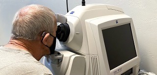
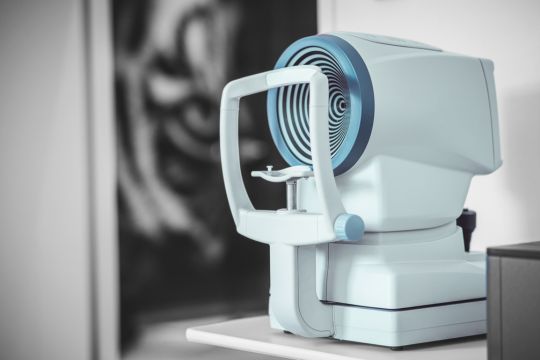
Optical Coherence Tomography (OCT)
An Optical Coherence Tomography scan (OCT scan) uses a scanning laser to analyze the layers of the retina and optic nerve for any signs of eye disease, similar to an CT scan of the eye. It works using light without radiation, and is essential for early diagnosis of glaucoma, macular degeneration and diabetic retinal disease.
With an OCT scan, doctors are provided with color-coded, cross-sectional images of the retina. These detailed images are revolutionizing early detection and treatment of eye conditions such as wet and dry age-related macular degeneration, glaucoma, retinal detachment and diabetic retinopathy.
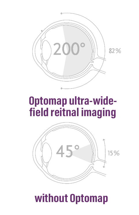
Optomap
Optomap detects 30% more than traditional dilation and is as simple as taking a photograph. The Optomap is a diagnostic tool that allows our doctors to view your retina by taking an ultra-wide view, high-resolution image of the back of your eye. It’s as simple as taking a photograph. Quite often using Optomap can replace traditional dilation.
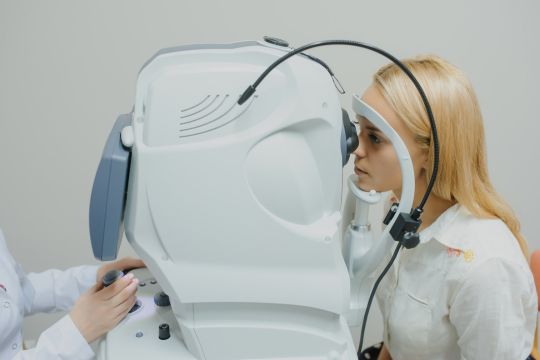
Corneal Topography
Corneal topography is a special photography technique that maps the surface of the clear, front window of the eye (the cornea). It gives our doctor a 3D map where they can find distortions in the curvature of the cornea, which is normally smooth. It also helps them monitor eye disease and plan for surgery. The scan only takes a few seconds, but it may need to be repeated a few times. Getting a corneal topography is painless, as nothing touches your eye during the scan.
When Is Corneal Topography Used?
- Scarring or injury of the cornea
- Growths can be monitored
- Astigmatism and keratoconus
- Contact lens fitting
- Refractive surgery
- Cataracts
- Corneal transplants
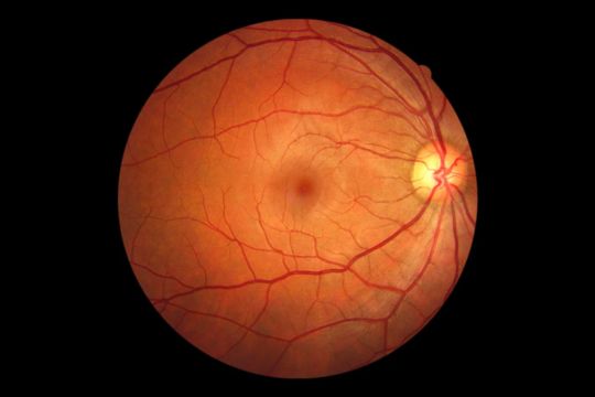
Digital Retinal Imaging
We use cutting-edge digital imaging technology to assess your eyes. Many eye diseases, if detected at an early stage, can be treated successfully without total loss of vision. Your retinal Images will be stored electronically. This gives your eye doctor a permanent record of the condition and state of your retina. Keeping these records are very important in assisting us to detect and measure any changes to your retina each time you have an examination, as many eye conditions, such as glaucoma, diabetic retinopathy and macular degeneration are diagnosed by detecting changes over time.
Retinal Imaging is a process carried out in seconds that is safe and painless. It aims to provide detailed images that other technologies might fail at and give instant high-resolution photos.
Our Imaging uses an eye-safe near-infrared light and doesn’t require the patient to prepare for it.
Dry Eye Treatments
